
Smart Optical Microscopy (SOM) Node is a multi-sited, multimodal Node including the Instituto de Salud Carlos III (ISCIII), the Centro Nacional de Investigaciones Oncológicas (CNIO), and Universidad Carlos III de Madrid (UC3M). The Node is designed to provide cutting-edge services in advanced fluorescence microscopy, with a specific focus on smart microscopy, high-throughput imaging, and AI-powered bioimage analysis to automate and adapt imaging workflows and support quantitative, scalable, and reproducible microscopy applications across a broad range of biomedical and translational research fields.
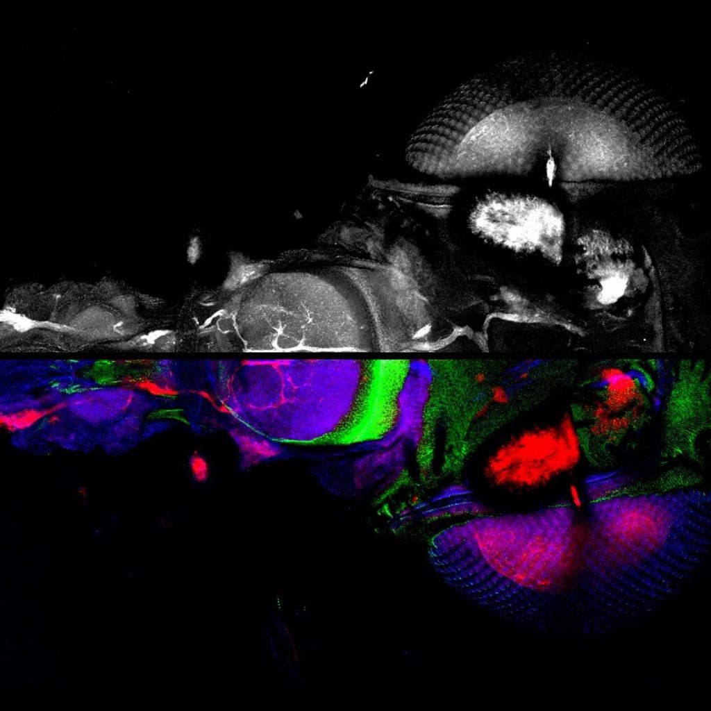
The Smart Optical Microscopy Node brings together advanced optical microscopy, automated imaging and artificial intelligence-based image analysis through the collaboration of three institutions: the Instituto de Salud Carlos III (ISCIII), the Spanish National Cancer Research Centre (CNIO), and Universidad Carlos III de Madrid (UC3M).
The Node provides integrated expertise across the imaging workflow, from experimental design and sample preparation to advanced image acquisition, quantitative analysis and data interpretation. Its capabilities support research in cell biology, oncology, tissue biology and biomedical research, with a particular focus on multidimensional imaging, high-content screening, multiplexed fluorescence imaging and AI-assisted bioimage analysis.
By combining complementary instrumentation and computational expertise across three institutions, SOM offers access to a broad portfolio of imaging technologies, automated platforms and advanced analytical workflows.
Offered Technologies
- Laser Scanning Confocal Microscopy (LSCM/CLSM)
Spectral confocal imaging, optical sectioning, multichannel fluorescence, 3D reconstruction and time-lapse imaging, with environmental control for live-cell experiments.
- Stimulated Emission Depletion Microscopy (STED)
STED super-resolution imaging, with reported resolution down to approximately 50 nm, for the visualization of subcellular structures beyond conventional diffraction-limited microscopy.
- Fluorescence Lifetime Imaging Microscopy (FLIM) and Gated Detection
Fluorescence lifetime measurements, lifetime-based contrast, gating and functional imaging, including FRET-related applications and live-cell experiments.
- Multiphoton Microscopy
Deep-tissue fluorescence imaging using near-infrared excitation, supporting imaging of optically thick biological samples and intravital applications.
- Widefield Fluorescence and Brightfield Microscopy
Automated and conventional widefield imaging, computational clearing, deconvolution, multichannel fluorescence and imaging of cells, tissues, slides and multiwell plates.
- Live-Cell Imaging and Time-Lapse Microscopy
Dynamic imaging of living cells under controlled temperature and CO₂ conditions, including long-term acquisition, microfluidics-compatible experiments and multidimensional time-lapse studies.
- High-Content Imaging and Screening (HCS)
Automated widefield and spinning-disk confocal imaging, robotic plate handling, automated incubation and high-throughput acquisition for quantitative 2D and 3D cellular assays.
- Autonomous and Automated Microscopy
Automated acquisition workflows, multi-position imaging, mosaic acquisition, autofocus and image-guided scanning for reproducible and scalable microscopy experiments.
- Whole-Slide and Tissue Imaging
Automated brightfield and fluorescence scanning of tissue sections, with multichannel acquisition, whole-slide digitization and support for quantitative tissue analysis.
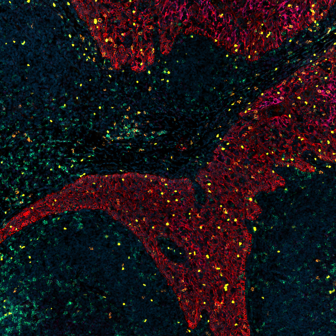
- Multiplexed Fluorescence Imaging and Cyclic Immunofluorescence
Multicolour tissue imaging and iterative staining workflows for the detection of multiple biological markers in the same sample, supporting spatially resolved tissue characterization.
- Holographic Tomography and Label-Free Imaging
Three-dimensional quantitative imaging based on cellular refractive index measurements, enabling label-free analysis of cell morphology and structure.
- Laser Photomanipulation and Targeted Ablation
Targeted laser manipulation of living cells, including localized cellular and subcellular ablation and DNA damage induction for functional experiments.
- AI-Assisted Bioimage Analysis and Deep Learning
GPU-enabled image processing, deep-learning-based segmentation, denoising and classification, custom model training, automated analysis pipelines and quantitative interpretation of 2D, 3D and time-lapse microscopy data.
- 3D and 4D Image Analysis
Multidimensional visualization, volume quantification, cellular and tissue segmentation, object tracking and analysis of dynamic biological processes across spatial and temporal dimensions.
- AI-Assisted Image Restoration
Image restoration and enhancement workflows designed to improve microscopy image quality under suboptimal acquisition conditions, including support for fast processing and reduced-light imaging strategies.
Additional Services offered by the Node
- Experimental Design and Sample Preparation
- Advice on experimental design and selection of suitable imaging approaches.
- Guidance on fluorophores, staining protocols and acquisition parameters.
- Support for live-cell, tissue and multidimensional imaging experiments.
- Advanced Image Acquisition
- Technical support for conventional and advanced optical microscopy.
- Assistance with automated acquisition, high-content screening and multiplexed imaging.
- Support for acquisition optimization and reproducible imaging workflows.
- Image Processing and Quantitative Analysis
- Image visualization, processing and quantitative analysis.
- 2D, 3D and 4D analysis of cells and tissues.
- AI-based segmentation, deep learning and custom analysis pipelines.
- Image restoration and analysis of high-content datasets.
- Computational Infrastructure and Data Management
- Access to image analysis workstations and institutional computational resources.
- GPU-enabled deep-learning workflows and local or remote model execution.
- Support for large image datasets and reproducible analysis pipelines.
- Training and Technical Consultancy
- Training in advanced microscopy and image acquisition.
- Training in quantitative image analysis, AI and deep learning.
- Support for workflow development and technology transfer.
Instrument available
- Spectral Confocal Microscopy and STED
Leica Stellaris and SP8 platforms provide spectral detection, fluorescence lifetime imaging and gated STED super-resolution. The portfolio also includes Leica SP5 systems and a Zeiss LSM990 confocal platform.
Institutions: ISCIII, CNIO and UC3M.
- Automated High-Content Imaging
Opera Phenix Plus and Operetta platforms combine widefield and spinning-disk confocal imaging with robotic plate handling and automated environmental control.
Institutions: CNIO and ISCIII.
- Whole-Slide Scanning and Multiplexed Tissue Imaging
Two Zeiss Axioscan 7 scanners support automated brightfield and fluorescence digitization of tissue slides, with multichannel acquisition for tissue-level studies.
Institutions: ISCIII and CNIO.
- Holographic Tomography
The Nanolive platform enables label-free 3D imaging and cellular refractive index measurements, with environmental control for live-cell experiments.
Institution: ISCIII.
- AI and Computational Microscopy
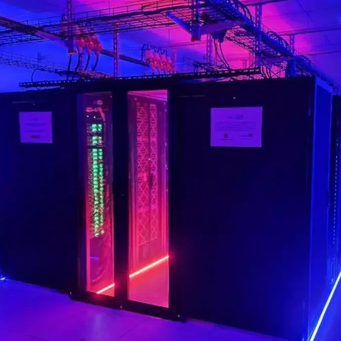
The Node combines dedicated image analysis workstations, institutional high-performance computing and software for deep-learning-based image analysis, including BiaPy and BioImage.IO BioEngine/Hypha.
Institutions: ISCIII, CNIO and UC3M.
- Spatial Biology Platform
Widefield epifluorescence platform with liquid handling for cyclic immunofluorescence staining, intended to support multiplexed tissue imaging and spatial biology workflows.
Institution: CNIO
Contact
Diego Megias – Instituto de Salud Carlos III
diego.megias@isciii.es
Isabel Peset – Centro Nacional de Investigaciones Oncológicas (CNIO)
ipeset@cnio.es
Arrate Muñoz – Universidad Carlos III de Madrid
mamunozb@ing.uc3m.es
Facilities